BioNMR
NMR aggregator & online community since 2003
BioNMR    
Learn or help to learn NMR - get free NMR books!
 

Go Back   BioNMR > NMR community > News from NMR blogs
Advanced Search
Home Forums Wiki NMR feeds Downloads Register Today's Posts


Jobs Groups Conferences Literature Pulse sequences Software forums Programs Sample preps Web resources BioNMR issues


Webservers
NMR processing:
MDD
NMR assignment:
Backbone:
Autoassign
MARS
UNIO Match
PINE
Side-chains:
UNIO ATNOS-Ascan
NOEs:
UNIO ATNOS-Candid
UNIO Candid
ASDP
Structure from NMR restraints:
Ab initio:
GeNMR
Cyana
XPLOR-NIH
ASDP
UNIO ATNOS-Candid
UNIO Candid
Fragment-based:
BMRB CS-Rosetta
Rosetta-NMR (Robetta)
Template-based:
GeNMR
I-TASSER
Refinement:
Amber
Structure from chemical shifts:
Fragment-based:
WeNMR CS-Rosetta
BMRB CS-Rosetta
Homology-based:
CS23D
Simshift
Torsion angles from chemical shifts:
Preditor
TALOS
Promega- Proline
Secondary structure from chemical shifts:
CSI (via RCI server)
TALOS
MICS caps, β-turns
d2D
PECAN
Flexibility from chemical shifts:
RCI
Interactions from chemical shifts:
HADDOCK
Chemical shifts re-referencing:
Shiftcor
UNIO Shiftinspector
LACS
CheckShift
RefDB
NMR model quality:
NOEs, other restraints:
PROSESS
PSVS
RPF scores
iCing
Chemical shifts:
PROSESS
CheShift2
Vasco
iCing
RDCs:
DC
Anisofit
Pseudocontact shifts:
Anisofit
Protein geomtery:
Resolution-by-Proxy
PROSESS
What-If
iCing
PSVS
MolProbity
SAVES2 or SAVES4
Vadar
Prosa
ProQ
MetaMQAPII
PSQS
Eval123D
STAN
Ramachandran Plot
Rampage
ERRAT
Verify_3D
Harmony
Quality Control Check
NMR spectrum prediction:
FANDAS
MestReS
V-NMR
Flexibility from structure:
Backbone S2
Methyl S2
B-factor
Molecular dynamics:
Gromacs
Amber
Antechamber
Chemical shifts prediction:
From structure:
Shiftx2
Sparta+
Camshift
CH3shift- Methyl
ArShift- Aromatic
ShiftS
Proshift
PPM
CheShift-2- Cα
From sequence:
Shifty
Camcoil
Poulsen_rc_CS
Disordered proteins:
MAXOCC
Format conversion & validation:
CCPN
From NMR-STAR 3.1
Validate NMR-STAR 3.1
NMR sample preparation:
Protein disorder:
DisMeta
Protein solubility:
camLILA
ccSOL
Camfold
camGroEL
Zyggregator
Isotope labeling:
UPLABEL
Solid-state NMR:
sedNMR


Reply
Thread Tools Search this Thread Rate Thread Display Modes
  #1  
Unread 01-19-2012, 09:19 PM
nmrlearner's Avatar
Senior Member
 
Join Date: Jan 2005
Posts: 23,175
Points: 193,617, Level: 100
Points: 193,617, Level: 100 Points: 193,617, Level: 100 Points: 193,617, Level: 100
Level up: 0%, 0 Points needed
Level up: 0% Level up: 0% Level up: 0%
Activity: 50.7%
Activity: 50.7% Activity: 50.7% Activity: 50.7%
Last Achievements
Award-Showcase
NMR Credits: 0
NMR Points: 193,617
Downloads: 0
Uploads: 0
Default Sorting Out NOE's for Exchanging Rotomers

Sorting Out NOE's for Exchanging Rotomers

2D NOESY spectra contain cross peaks from both NOE interactions and peaks due to rotomers in slow exchange with one another on the NMR tine scale. For small molecules, the cross peaks resulting from slowly exchanging rotomers are of the same sign as the diagonal peaks. The NOE cross peaks, on the other hand, are of opposite sign compared to the diagonal peaks. When both types of correlations are present there may be more NOE correlations than expected. What follows is an example of this. The figure below shows a color coded chemical structure of a ruthenium complex with a color coded partial 1H NMR spectrum.
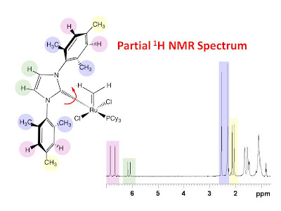
It is obvious from the NMR spectrum that all of the signals from the color coded protons are doubled in the spectrum. One possible explanation for this is that there is a slow rotation about the ruthenium carbon bond indicated with the red curly arrow allowing for two possible nonequivalent rotomers. This is confirmed with the 2D 1H NOESY spectrum shown in the figure below with a 0.9 second mixing time. The spectrum clearly shows exchange peaks between corresponding pairs of 1H signals from each rotomer.The interesting thing to note from the NOESY spectrum is that each aromatic proton (pink) from a single rotomer shows NOE correlations to the methyl groups (blue and yellow) of both rotomers - not just those from a single rotomer. With this data, it is not possible to assign the subspectrum of a single rotomer. Presumably, the assignment could be made by collecting a 2D NOESY spectrum at low temperature where the rotation was completely frozen out or by collecting a 2D NOESY spectrum with a very short mixing time where the rotation would be limited. The problem with the former approach is that the solvent may freeze at a temperature too high to stop the bond rotation. The problem with the latter approach is that the NOE's would be much reduced due to the short mixing time and collecting a 2D data set with sufficient signal to noise ratio would take a great deal of time. Another approach is to collect selective 1D gradient NOESY spectra with selective excitation of the aromatic proton from each rotomer individually. These data are shown in the figure below for two different mixing times.Each spectrum is displayed in two parts. The left-hand panel is the aromatic region with the selectively excited resonance colored red and the right-hand panel is the aliphatic region showing the NOE correlations to the methyl groups. From the spectra collected with a 2 second mixing time, one can see that the selective excitation is no longer selective due to bond rotation during the long mixing time. One can see inverted peaks for the aromatic protons of both rotomers despite the fact that the 1H signal of only one rotomer was selectively excited. Furthermore, NOEs to the methyl signals from both rotomers are present. The spectra collected with only a 0.2 second mixing time, on the other hand, show very selective excitation. The time scale of the bond rotation is obviously longer than the 0.2 second mixing time. The spectra show only the NOEs between the selectively excited aromatic proton and the methyl groups from a single rotomer. The NOEs build up fast enough to be observed during the 0.2 second mixing time before rotation occurs. These data allow for the assignment of signals from each of the rotomers.

Thank you to Justin Lummiss of Dr. Fogg's research group for aharing this interesting system.


Source: University of Ottawa NMR Facility Blog
Reply With Quote


Did you find this post helpful? Yes | No

Reply
Similar Threads
Thread Thread Starter Forum Replies Last Post
Using the water signal to detect invisible exchanging protons in the catalytic triad of a serine protease
Using the water signal to detect invisible exchanging protons in the catalytic triad of a serine protease Abstract Chemical Exchange Saturation Transfer (CEST) is an MRI approach that can indirectly detect exchange broadened protons that are invisible in traditional NMR spectra. We modified the CEST pulse sequence for use on high-resolution spectrometers and developed a quantitative approach for measuring exchange rates based upon CEST spectra. This new methodology was applied to the rapidly exchanging Hδ1 and Hε2 protons of His57 in the catalytic triad of bovine chymotrypsinogen-A...
nmrlearner Journal club 0 07-25-2011 11:01 AM
[NMR paper] Sorting signals from protein NMR spectra: SPI, a Bayesian protocol for uncovering spi
Sorting signals from protein NMR spectra: SPI, a Bayesian protocol for uncovering spin systems. Related Articles Sorting signals from protein NMR spectra: SPI, a Bayesian protocol for uncovering spin systems. J Biomol NMR. 2002 Nov;24(3):203-13 Authors: Grishaev A, Llinás M Grouping of spectral peaks into J-connected spin systems is essential in the analysis of macromolecular NMR data as it provides the basis for disentangling chemical shift degeneracies. It is a mandatory step before resonance and NOESY cross-peak identities can be...
nmrlearner Journal club 0 11-24-2010 08:58 PM


Posting Rules
You may not post new threads
You may not post replies
You may not post attachments
You may not edit your posts

BB code is On
Smilies are On
[IMG] code is On
HTML code is Off
Trackbacks are Off
Pingbacks are Off
Refbacks are Off


BioNMR advertisements to pay for website hosting and domain registration. Nobody does it for us.


Powered by vBulletin® Version 3.7.3
Copyright ©2000 - 2024, Jelsoft Enterprises Ltd.
Copyright, BioNMR.com, 2003-2013
Search Engine Friendly URLs by vBSEO 3.6.0

All times are GMT. The time now is 08:29 AM.


Map