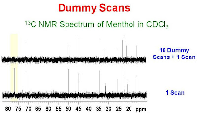
Dummy scans (DS, Bruker) or steady state scans (SS, Varian) are scans taken in an NMR acquisition before the receiver is turned on and data are collected. Each dummy scan contains all of the rf pulses, delays and gradients used in the pulse program; the only difference is that the receiver is not turned on to collect data. Dummy scans are typically used to ensure that a spin system is in a steady state before data are collected. For example, One may collect a spectrum using a relaxation delay short with respect to the T1's of some of the resonances in the spectrum. The first scan may find the system at equilibrium, as the sample may have sat in the magnet for a few minutes while the probe was being tuned and matched or the magnet was being shimmed. The second and subsequent scans will find the system not at equilibrium, as the system has been perturbed by the rf pulses of the preceding scan(s) and not allowed to fully relax. The data from the first and seccond scan are therefore not the same. After several scans, although not at equilibrium, the system will be in a steady state before each additional scan and therefore each subsequent scan will collect similar data. An example of the use of dummy scans is shown in the figure below. The bottom trace shows a single scan 13C NMR spectrum of a concentrated solution of menthol in CDCl3 after the sample had sat in the magnet for a minute or so. The top trace shows a similar single scan acquisition preceded by 16 dummy scans. The relaxation delay was set to 1 second and the acquisition time for the FID was 1 second. 90° pulses were used in both spectra. One can see that some of the resonances in the top spectrum are attenuated in comparison to the bottom spectrum. The 13C signal from the CDCl3 (which has a long T1) is missing in the spectrum acquired with dummy scans. The dummy scans have selectively presaturated the solvent.
peak intensity vs number of scans
Hi,
I have a question regarding how the peak intensities change when with increase in number of scans when the data is collected on a Varian NMR spectrometer. Suppose I collect a 15N-1H HSQC spectra with 8 scans and then collect the same spectra with 16 scans, would the peak intensities be double in the 16 scan spectra as compared to the 8 scan spectra? Does anyone know how the varian spectrometer scales the intensities according to the number of scans? Any suggestion or reply to my query will be extremely useful and greatly appreciated.
Thanks.
aish1982
NMR software
0
08-08-2008 11:56 PM
peak intensity vs number of scans
Hi,
I have a question regarding how the peak intensities change when with increase in number of scans when the data is collected on a Varian NMR spectrometer. Suppose I collect a 15N-1H HSQC spectra with 8 scans and then collect the same spectra with 16 scans, would the peak intensities be double in the 16 scan spectra as compared to the 8 scan spectra? Does anyone know how the varian spectrometer scales the intensities according to the number of scans? Any suggestion or reply to my query will be extremely useful and greatly appreciated.
Thanks.


 The bottom trace shows a single scan 13C NMR spectrum of a concentrated solution of menthol in CDCl3 after the sample had sat in the magnet for a minute or so. The top trace shows a similar single scan acquisition preceded by 16 dummy scans. The relaxation delay was set to 1 second and the acquisition time for the FID was 1 second. 90° pulses were used in both spectra. One can see that some of the resonances in the top spectrum are attenuated in comparison to the bottom spectrum. The 13C signal from the CDCl3 (which has a long T1) is missing in the spectrum acquired with dummy scans. The dummy scans have selectively presaturated the solvent.


 Linear Mode

